Compiled from clinical pathology references. Medically reviewed by Dr Cristian Dunker, Principal Dentist, ArtSmiles Cosmetic Dentistry.
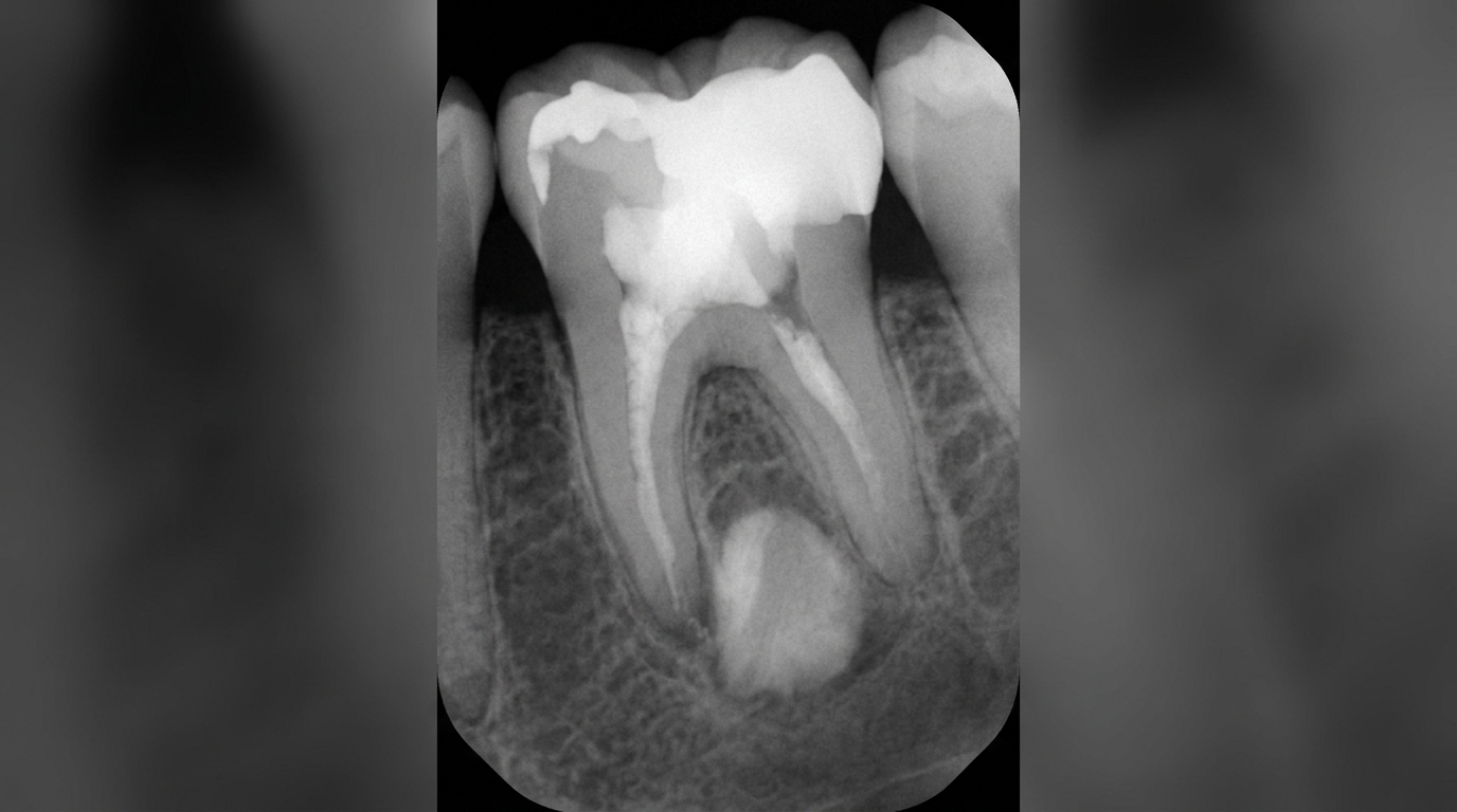
A small, well-defined patch of denser, whiter-than-usual bone next to the root of a back tooth on a dental X-ray often catches the eye. The name for this finding is condensing osteitis, also known as focal sclerosing osteomyelitis. The good news is that, in itself, it is harmless. It tells us about the tooth above it, not about a problem in the bone.
This article from the team at ArtSmiles, reviewed by Dr Cristian Dunker, explains what condensing osteitis is, why it forms, and what (if anything) needs to be done.
What is it?
Condensing osteitis is a localised area of increased bone density at the tip of a tooth root. On a dental X-ray it shows up as a radiopaque (whiter than the surrounding bone) area, sometimes round, sometimes irregular, blending into the bone at its margins.
Microscopically, it represents bone that has laid down extra material in response to chronic, low-grade inflammation in the dental nerve above. It is not infection of bone in the way that osteomyelitis is, there is no pus, no bacterial invasion of the bone marrow, no destruction. It is a reactive change, similar in spirit to a callus on the skin.
Who tends to get it?
Condensing osteitis can be found in patients of any age but is most common in:
Adults under 40 years old.
Patients with a deep filling, large cavity or root canal-treated tooth at the site.
Patients with a history of repeated dental work on the affected tooth (deep cavities, replaced fillings, large restorations).
People with a tooth that has been mildly tender or sensitive over months or years, sometimes too mildly to prompt a visit.
It is most often seen at the lower first molar, although it can occur at any tooth.
What causes it?
Condensing osteitis develops in response to low-grade, chronic inflammation in the dental pulp, the nerve and blood vessels inside the tooth. Common triggers include:
A deep cavity that has been close to the nerve for a long time.
A large filling with low-level pulp irritation.
A cracked tooth with chronic micro-injury to the pulp.
Previous trauma that reduced the tooth's blood supply over years.
A non-vital tooth (a tooth whose nerve has died) that is slowly leaking inflammatory mediators into the surrounding bone.
The bone immediately around the root tip responds to these chronic signals by laying down more bone, producing the characteristic dense area.
How does it develop?
The course is gradual:
The tooth nerve is irritated, often without much pain.
Low-grade inflammatory signals reach the surrounding bone over months to years.
Bone-forming cells (osteoblasts) lay down extra bone in the area.
A dense, well-defined area builds up at the root tip.
The change is found incidentally on a dental X-ray.
After the tooth is treated (root canal, restoration or extraction), the dense bone often persists for years; some lesions slowly regress.
What might you notice?
In most cases, nothing. Condensing osteitis itself is silent. The tooth above it may have:
No symptoms at all, the inflammation has been so low-grade that the patient has noticed nothing.
Mild sensitivity to hot, cold or sweet foods.
Discomfort on biting that comes and goes.
A slight ache that has been ignored for a long time.
A history of past sensitivity that resolved.
Many patients are told about condensing osteitis only when their dentist points it out on a routine X-ray.
What happens at the dentist?
When condensing osteitis is found at ArtSmiles, the visit usually involves:
A look at the X-ray together with you, explaining what we see.
A clinical examination of the tooth above the dense area, checking for cracks, defective fillings or large cavities.
Vitality testing (a quick test to check whether the nerve inside the tooth is still alive) of the tooth to see whether the nerve is still alive.
Bite testing and percussion (gentle tapping on the tooth to check tenderness and bite response) to see how the tooth responds.
A management decision:
Tooth alive and asymptomatic: monitor at routine reviews.
Tooth with reversible inflammation: address the cause (replace a faulty filling, treat decay).
Tooth with non-vital nerve: root canal therapy.
Tooth that cannot be saved: extraction.
Post-treatment review with imaging at 6 to 12 months.
Is this serious?
Condensing osteitis is not serious in itself. The reasons it deserves attention are:
It marks a tooth that has had inflammation, which often needs treatment.
A non-vital tooth can lead to problems later if not treated.
Some other conditions can produce dense bone areas and need to be excluded.
It is not cancer, not infection of bone in the dangerous sense, and does not spread.
Could it be something else?
Other dense bone changes can look similar. Considerations include:
Idiopathic osteosclerosis (dense bone island). A perfectly normal incidental finding with no link to a tooth.
Cementoblastoma. A benign tumour fused to a tooth root.
Osteoma. A benign bone tumour.
Hypercementosis. Excess cementum (the thin layer covering the tooth root) on the root surface, often in older adults.
Periapical cemento-osseous dysplasia. Mixed dense and dark areas around tooth roots.
Chronic sclerosing osteomyelitis. A more diffuse, infection-related dense bone change.
A clinical examination and the relationship of the dense area to a tooth usually clarify which one it is.
How is it treated?
Condensing osteitis itself does not need treatment. The treatment is directed at the tooth responsible:
Routine monitoring if the tooth is healthy and asymptomatic.
Replacing a faulty filling or treating a cavity if the nerve is irritated but recoverable.
Root canal therapy if the nerve is no longer vital.
Extraction if the tooth is not restorable.
Follow-up imaging at 6 to 12 months to ensure the area is stable or improving.
Some condensing osteitis areas remain visible on X-rays for many years even after the original tooth has been treated or removed. This is normal and does not indicate ongoing disease.
What's the long-term outlook?
The long-term outlook is excellent. Once the underlying tooth has been managed, either kept healthy, root-canal-treated or extracted, the area of dense bone is no longer a clinical concern, even if it remains visible on later X-rays. Regular check-ups are all that is needed for ongoing reassurance.
If condensing osteitis has been mentioned to you on a recent X-ray and you are unsure what it means, please book a visit so we can review the film with you and make a clear plan together.
A note on this article
This article is for educational purposes only and does not constitute a clinical diagnosis. Please consult a registered dental practitioner for assessment and treatment advice.
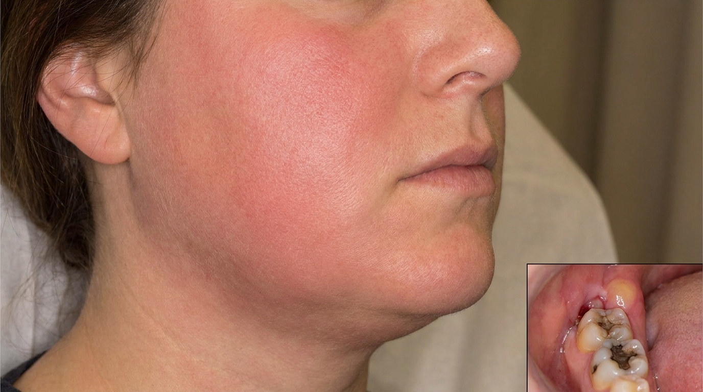
The cover image above is an AI-generated illustration based on the most common visible features of this condition described in clinical pathology references. It is not a photograph of a real case and should not be used to diagnose or rule out the condition in your own situation. If you are concerned about something you have noticed, please book an assessment with a registered dental practitioner.
References
Neville, B. W., Damm, D. D., Allen, C. M., & Chi, A. C. (2016). Oral and maxillofacial pathology (4th ed., Ch. 3: Pulpal and Periapical Disease, Condensing Osteitis). Elsevier.
Cawson, R. A., & Odell, E. W. (2017). Cawson's essentials of oral pathology and oral medicine (8th ed., Ch. 6: Periapical Inflammatory Disease). Elsevier.
Regezi, J. A., Sciubba, J. J., & Jordan, R. C. K. (2017). Oral pathology: clinical pathologic correlations (7th ed., Ch. 9: Bone Lesions). Elsevier.
Frequently asked questions
What is condensing osteitis?
Condensing osteitis (also called focal sclerosing osteomyelitis) is a localised thickening of the jawbone around the root of a tooth with chronic low-grade infection or irritation, usually from a long-standing pulp problem. The body lays down extra bone instead of breaking it down, producing the bright white patch seen on an X-ray.
Is condensing osteitis dangerous?
No. It is a benign reactive change in the bone, not a tumour and not a cancer. It is mainly important as a sign that something is going on with the nearby tooth: the affected tooth often has an inflamed or dead pulp that needs treatment.
How is condensing osteitis treated?
Treatment focuses on the source tooth. Options include root canal treatment to remove the infected pulp, or extraction if the tooth cannot be saved. In many cases, the bone changes slowly return to normal over months after the underlying cause is resolved.
Will the white spot on my X-ray go away?
Often yes. Once the source of irritation in the tooth is treated, the dense bone usually remodels and partially or fully fades on follow-up X-rays. A small residual area (sometimes called an osteosclerotic scar) may persist as a permanent mark, but it is harmless.