Compiled from clinical pathology references. Medically reviewed by Dr Cristian Dunker , Principal Dentist, ArtSmiles Cosmetic Dentistry.
Quick summary
Also called | Salivary gland stones, salivary calculi, sialolith |
How urgent? | 🟡 Worth assessing, usually not dangerous, but can cause repeated pain, swelling and infection until the stone is removed |
Common or rare? | The most common cause of salivary gland obstruction |
Who it affects | Mostly adults, particularly men, with about twice as many cases in men as women; most commonly affects the submandibular gland (around 80% of cases) |
Who treats it | General dentist for diagnosis and milder cases, with referral to an oral and maxillofacial surgeon, ENT surgeon or interventional radiologist when more involved treatment is needed |
Based on | Cawson, Laskaris, with cross-references in Neville |
What is it?
Sialolithiasis is the formation of one or more stones inside a salivary gland or its duct. The stones, also called sialoliths or salivary calculi, are usually made of calcium salts deposited around a small organic core within the saliva. As they grow, they obstruct the flow of saliva from the gland into the mouth. Because saliva production rises sharply at the smell, sight or first taste of food, the classic symptom is swelling and pain in the gland at meal times, easing once the meal is finished. Sialolithiasis is the most common cause of salivary gland obstruction and, although the symptoms can be uncomfortable, the condition is usually well managed once the stone is identified.
Who tends to get it?
The textbooks describe a fairly consistent profile:
Adults are mainly affected, with men involved roughly twice as often as women.
The submandibular gland (the gland under the lower jaw) accounts for about 80% of all cases.
The parotid gland (the large gland in front of the ear) accounts for around 6% of cases.
The sublingual and minor salivary glands account for around 2% of cases.
Usually unilateral, one side at a time.
People with a history of dehydration, low fluid intake, dry mouth or chronic salivary gland inflammation are at higher risk.
The submandibular gland is particularly prone to stones because its saliva is more mucous and its duct (Wharton's duct (the duct that drains saliva from the submandibular gland into the floor of the mouth)) runs uphill against gravity from the floor of the mouth into the gland.
What causes it?
The textbooks describe several factors that contribute to stone formation:
Stagnation of saliva in the duct, for example, in dehydration, reduced flow, or after a small disturbance of normal flow.
Deposition of calcium salts around an organic nucleus (often debris, bacteria, or desquamated cells from the duct lining).
Layered (lamellar) growth, stones build up over weeks to months in concentric rings, gradually enlarging.
Composition of saliva, the higher mucin content of submandibular saliva and its slightly higher pH favour calcium salt deposition.
Local infection or inflammation, chronic sialadenitis can both cause and be aggravated by stones, in a self-perpetuating cycle.
There is no strong evidence that diet, calcium intake, or specific systemic conditions cause sialolithiasis, although patients with a history of kidney stones may be at slightly higher risk in some series.
How does it develop?
Once a small concretion has formed, it gradually enlarges as more calcium salts are deposited around it. The roughness of the stone irritates the duct lining, causing the lining to thicken and sometimes undergo squamous metaplasia (cell-type change in the duct lining). An adherent layer of bacteria can grow on the surface of the stone, which together with the obstruction often triggers inflammation and fibrosis around the duct (chronic sialadenitis).
When the patient eats, or even smells food, the gland produces a sudden surge of saliva. The stone partially blocks the duct, so saliva backs up and the gland swells. The pressure stretches the gland capsule and produces the characteristic meal-time pain. After the meal, saliva flow drops, the pressure eases, and the swelling gradually settles. Over months and years, repeated obstruction can damage the gland, leading to chronic sialadenitis, fibrosis and reduced saliva production from that gland.
What might you notice?
What it looks like
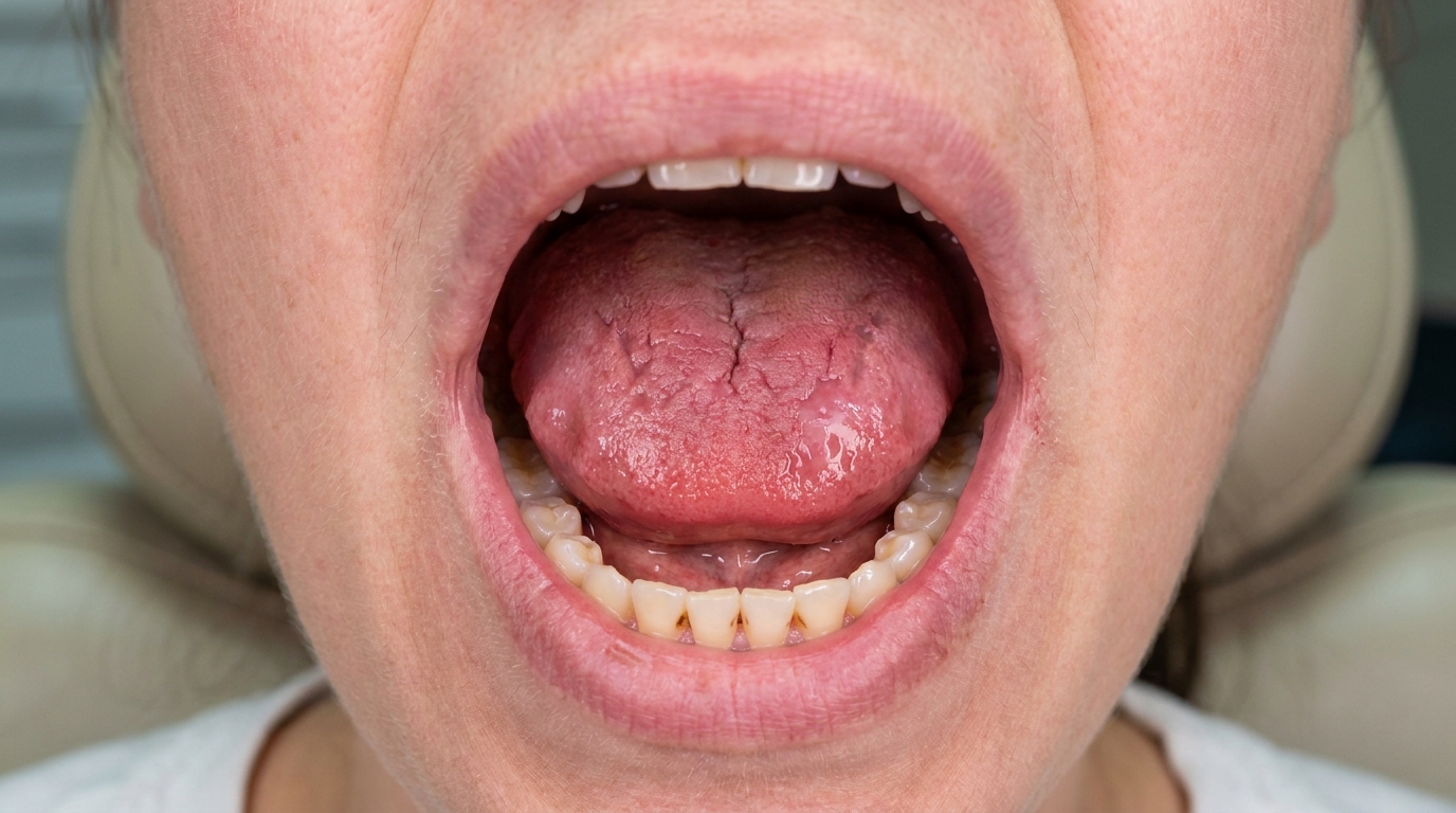
Most stones are inside the duct or gland and not visible from outside. Visible signs may include:
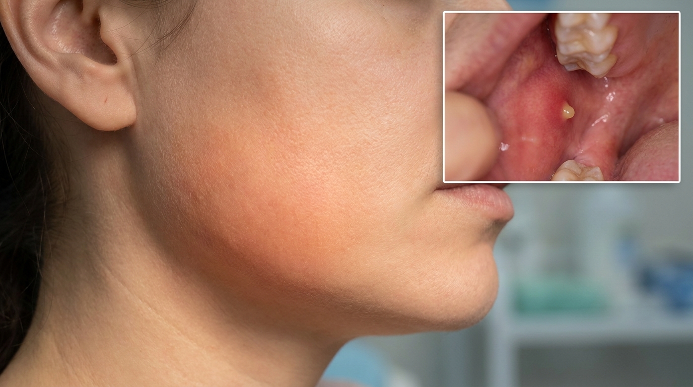
A swelling under the jaw or in the cheek, particularly at meal times, that gradually settles afterwards.
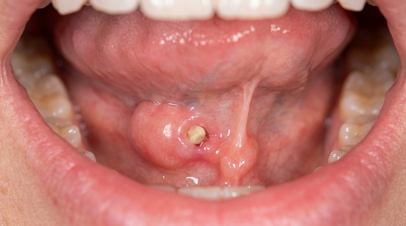
A small yellow or white nodule visible at the duct opening under the tongue or inside the cheek if the stone has migrated forward.
Swelling and redness of the floor of the mouth near the duct opening if there is associated infection.
Pus at the duct opening if the gland is acutely infected.
What it feels like
The classic symptoms described in the textbooks include:
Pain on the side of the face or under the jaw that develops with the smell, sight or taste of food and eases after the meal.
A tender swelling that comes and goes with eating.
A constant tender swelling if the gland has become infected (sialadenitis).
A bad or salty taste in the mouth from leaking saliva or pus.
Sometimes no symptoms at all, particularly with smaller stones that pass forward and can be felt in the mouth or seen by chance on a routine X-ray.
Importantly, the textbooks specifically note that sialolithiasis itself does not cause persistent dry mouth. Patients with one obstructed gland still have many other functioning salivary glands, so overall saliva production stays in the normal range.
What an X-ray might show
Imaging is the most useful tool for confirming a salivary stone:
A plain dental X-ray (often a lower occlusal film) usually shows submandibular stones, since most of them are radiopaque (shows up on X-rays as a white shape).
A panoramic X-ray can also show stones, though smaller ones may be missed.
About 40% of parotid stones and 20% of submandibular stones are not radiopaque, in which case a sialogram (special X-ray after dye injection into the duct) or ultrasound is used.
Cone-beam CT or conventional CT scans give excellent detail for planning surgery in difficult cases.
Sialendoscopy uses a tiny camera passed up the duct, both diagnostic and therapeutic, and is increasingly used in specialist centres.
What happens at the dentist?
A patient with mealtime salivary swelling is most often seen at a routine dental check-up and clean at ArtSmiles or at a problem-focused appointment. The dentist will typically:
Take a careful history, asking about the timing, side, severity and pattern of swelling, and any history of dehydration, dry mouth or previous infections.
Examine the gland externally and palpate it bimanually (with one finger inside the mouth and one outside) (one finger in the mouth, one outside) to feel for stones in the floor of the mouth.
Inspect the duct opening for swelling, pus or a visible stone.
Massage the gland and observe whether the saliva flow is normal, scant, or contains pus.
Take an appropriate X-ray to look for the stone, often a lower occlusal view for the submandibular gland.
Refer to an oral and maxillofacial surgeon, ENT surgeon or interventional radiologist when more advanced imaging or treatment is needed.
Manage any acute infection with antibiotics and supportive care.
Is this serious?
🟡 Sialolithiasis itself is rarely dangerous, but if a stone is left untreated the gland can be repeatedly damaged by obstruction and infection, and may eventually need to be removed. The most important reason to act early is to relieve symptoms and protect the gland's long-term function. Acute infection on top of an obstructed gland can cause significant swelling, fever and discomfort, and occasionally requires hospital care.
If you have noticed pain or swelling under the jaw or in the cheek that comes on with eating and settles afterwards, or if a swelling in that area is becoming larger, redder or more painful, it is worth booking an assessment so the cause can be identified and the right treatment planned.
Could it be something else?
Several conditions can produce similar swellings around the salivary glands. The textbooks list these as the main differentials:
Acute bacterial sialadenitis, infection of the gland with pain, swelling and pus from the duct, often without an underlying stone.
Chronic sialadenitis, repeated low-grade inflammation, often without a single identifiable stone, leading to gland shrinkage and reduced flow.
Mumps, a viral infection (paramyxovirus) that classically causes painful swelling of one or both parotid glands, more common in children.
Salivary gland tumour, a slowly enlarging, persistent mass that does not fluctuate with meals; needs imaging and biopsy.
Sjögren syndrome, an autoimmune disease that causes generalised salivary gland involvement and dry mouth.
Ranula, a salivary cyst in the floor of the mouth that can be confused with sublingual stones.
Xerostomia, a different problem (reduced saliva flow overall) that can sometimes be misattributed to a single gland.
Lymphadenitis, inflamed lymph nodes near the gland that can mimic gland swelling.
How is it treated?
Treatment depends on the size and position of the stone, whether the gland is infected, and how much damage has already occurred. The textbooks describe a clear hierarchy from least to most invasive.
At-home measures and habits:
Stay well hydrated, sip water regularly through the day.
Sialagogues, sucking on a sugar-free sour lolly, lemon drop, or chewing sugar-free gum stimulates saliva flow and may help dislodge a smaller stone.
Warm compresses over the affected gland, with gentle external massage, can help in some cases.
Avoid medications that worsen dry mouth where possible, in discussion with your GP.
Stop or reduce smoking, since it can worsen overall saliva quality.
Professional steps your dentist may consider:
Manipulation and milking of a stone forward through the duct, particularly when it is close to the duct opening. Sometimes this alone is all that is needed.
Lithotripsy, using ultrasonic shock waves applied externally to break the stone into smaller pieces that can pass naturally.
Basket retrieval, a fine wire basket passed up the duct under fluoroscopic (live X-ray guidance) guidance to capture and remove the stone.
Sialendoscopy, a tiny camera passed up the duct, often combined with laser or basket disruption of the stone.
Surgical opening of the duct, under local anaesthetic, an incision is made along the duct to release a stone too large to pass naturally. The duct is left to heal, often with the margins sutured to the surrounding mucosa to prevent scarring.
Surgical removal of the gland (sialadenectomy), reserved for stones inside the gland itself or for glands that have been severely damaged by recurrent infection and fibrosis.
Antibiotics for any acute bacterial infection, typically based on culture and sensitivity testing.
A patient-centred approach matters here. Recurring meal-time swelling can be confusing and frustrating, particularly when the cause is not yet known. Honest, unhurried explanation of what is happening, what imaging is needed, and which option fits best is itself part of effective care, values that sit at the heart of our clinical philosophy.
What's the long-term outlook?
The outlook is generally very good. Most stones can be removed by minimally invasive techniques, and the affected gland often recovers normal function over weeks to months, even after repeated attacks of chronic sialadenitis. Stones can recur, particularly in patients with persistent risk factors such as dehydration or dry mouth, so attention to hydration and salivary flow remains important after treatment. In a small minority of patients with severely damaged glands, removal of the gland is needed, but the remaining salivary glands generally compensate well and overall mouth function stays normal.
A note on this article
This article is for educational purposes only and does not constitute a clinical diagnosis. Please consult a registered dental practitioner for assessment and treatment advice.
The cover image above is an AI-generated illustration based on the most common visible features of this condition described in clinical pathology references. It is not a photograph of a real case and should not be used to diagnose or rule out the condition in your own situation. If you are concerned about something you have noticed, please book an assessment with a registered dental practitioner.
References
Cawson, R. A., & Odell, E. W. (2017). Cawson's essentials of oral pathology and oral medicine (8th ed.). Elsevier. Chapter 18, Neoplastic and Non-Neoplastic Diseases of Salivary Glands: Salivary calculi, with key features in Box 18.1, lithotripsy (ultrasonic shock-wave treatment to break stones into smaller pieces) and basket retrieval, pp. 291 to 292.
Laskaris, G. Pocket atlas of oral diseases. Thieme. Chapter 34, Other Salivary Gland Disorders: Sialolithiasis, p. 332.
Neville, B. W., Damm, D. D., Allen, C. M., & Chi, A. C. (2023). Oral and maxillofacial pathology (5th ed.). Elsevier. Chapter on Salivary Gland Pathology: salivary calculi as the most common cause of salivary obstruction.