Compiled from clinical pathology references. Medically reviewed by Dr Cristian Dunker , Principal Dentist, ArtSmiles Cosmetic Dentistry.
Quick summary
Also called | Suppurative parotitis (when the parotid (the large salivary gland in front of the ear) gland is involved), acute parotitis |
How urgent? | 🔴 Important, needs prompt treatment with antibiotics and fluids; in debilitated or dehydrated patients, the infection can spread and become serious |
Common or rare? | Uncommon overall, but more frequent after surgery, in dehydrated patients, in those with chronic dry mouth, and in elderly or unwell adults |
Who it affects | Most often adults, particularly older or unwell patients with reduced salivary flow; the parotid gland is most commonly involved, with bilateral disease in 10-25% of cases |
Who treats it | General dentist or GP for early assessment, with referral to an oral and maxillofacial surgeon, ENT surgeon or hospital team if severe or not responding |
Based on | Cawson, Neville, with cross-references in Regezi |
What is it?
Acute bacterial sialadenitis (inflammation of a salivary gland) is an infection of a salivary gland by bacteria that have travelled up the duct from the mouth. The gland becomes painful, swollen and tender, and pus can often be expressed from the duct opening when the gland is gently massaged. The textbooks describe the parotid gland, the large gland in front of the ear, as the most commonly affected, where the condition is sometimes called suppurative parotitis. The submandibular (the salivary gland below the jaw) and sublingual glands can also be involved. Most cases are treated effectively with antibiotics and rehydration, but in debilitated patients the infection can be serious, with reported mortality rates of up to 20-50% in some severely unwell groups.
Who tends to get it?
The textbooks describe a fairly recognisable risk profile:
Older or debilitated adults, particularly after surgery (sometimes called "surgical mumps"), serious illness, or prolonged hospital stays.
Patients with reduced saliva flow, from dehydration, medications that cause dry mouth, Sjögren syndrome, or radiotherapy to the head and neck.
People with salivary gland stones, the most common single cause of obstruction that triggers infection.
People with congenital duct strictures or external compression of the duct (for example, by a salivary tumour).
People taking medications that reduce saliva, including anticholinergics, some antidepressants, antihistamines, and certain anti-Parkinson drugs.
Children with juvenile recurrent parotitis, a separate but related condition with repeated non-suppurative parotid swelling that usually resolves around puberty.
Patients on tricyclic antidepressants are noted by Cawson as an uncommon group.
The textbooks specifically note that with modern post-operative care, the classical "post-surgical parotitis" is much less common than it once was, and most cases now arise in ambulant patients with severe dry mouth.
What causes it?
The textbooks describe a fairly clear chain of events:
Reduced saliva flow allows bacteria to travel back up the duct (retrograde spread) instead of being flushed out.
Bacterial colonisation then triggers infection of the gland tissue.
Common organisms, most cases in the community are caused by Staphylococcus aureus or Streptococcus species.
Hospital-acquired infections, most often S. aureus but also a wider range of bacteria including Eikenella corrodens, Escherichia coli, Fusobacterium, Haemophilus influenzae, Klebsiella, Prevotella, Proteus and Pseudomonas.
Underlying obstruction, most commonly a salivary gland stone, but also congenital strictures, mucous plugs or tumour compression, sets up the conditions for infection.
Other contributing factors, dehydration, debilitation, immune suppression, and certain medications.
How does it develop?
When saliva flow falls, the protective washing action of saliva is lost and bacteria from the mouth can multiply within the duct system. As bacteria spread up the duct, they invade the surrounding gland tissue, triggering an acute inflammatory response. Neutrophils accumulate in the ducts and acini (the small clusters of saliva-producing cells), and pus develops within the gland. The pressure of the inflammation, the swelling of the tissues, and the obstruction of the duct combine to produce the characteristic painful, tender, hot swelling. If the infection is not treated, an abscess can form, occasionally rupturing through the skin or into the mouth.
What might you notice?
What it looks like
The classic clinical picture includes:
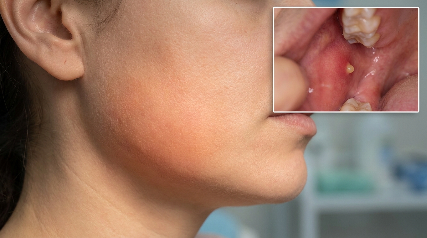
A painful swelling of the affected gland, most often in front of the ear (parotid) or under the jaw (submandibular).
Warm, red and tender overlying skin.
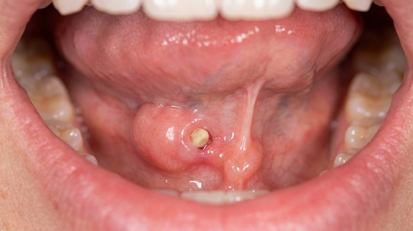
Pus visible at the duct opening when the gland is gently massaged from outside.
Limited mouth opening (trismus) when the infection is severe.
Bilateral involvement in about 10-25% of parotid cases, both sides at once.
What it feels like
Symptoms typically include:
Constant, severe, throbbing pain in the affected gland.
Tenderness when the area is gently touched.
A bad or salty taste in the mouth from leaking pus.
Reduced mouth opening because the muscles nearby are inflamed.
Low-grade fever and a sense of being unwell.
Worsening pain when eating, although the pain is usually constant rather than only at meal times, distinguishing it from the meal-related pain of a simple stone.
What an X-ray might show
Imaging is helpful to look for an underlying cause and to assess the gland:
A panoramic dental X-ray is often the first investigation, looking for a salivary gland stone.
Ultrasound is widely used and can show gland enlargement, ductal dilatation and abscess formation.
CT scan with contrast is reserved for more serious cases or when an abscess is suspected.
MRI may be used in selected cases, particularly if a tumour is suspected.
What happens at the dentist?
A patient with a hot, painful salivary swelling is most often seen at a problem-focused appointment rather than at a routine dental check-up and clean at ArtSmiles. The dentist will typically:
Take a careful history, recent illness, surgery, medications, fluid intake, history of dry mouth or salivary stones, and the timing of the swelling.
Examine the gland externally and look for skin redness and warmth.
Inspect the duct opening in the mouth and gently massage the gland to look for pus.
Take a swab of any pus for bacterial culture and sensitivity testing.
Take a panoramic X-ray as a screening tool for any underlying salivary stone.
Start antibiotic therapy appropriate for the likely organisms while awaiting culture results.
Recommend rehydration, mild analgesia and saliva-stimulating measures.
Refer to a hospital team or specialist if the patient is systemically unwell, dehydrated, immunocompromised, or shows signs of an abscess.
Is this serious?
🔴 In most healthy adults, acute bacterial sialadenitis is uncomfortable but not dangerous, and responds quickly to antibiotics and fluids. In debilitated, elderly or immunocompromised patients, however, the infection can spread, sepsis can develop, and serious complications are possible. The textbooks note reported mortality of 20-50% in severely debilitated patients with overwhelming infection. Prompt treatment, careful follow-up and a low threshold for hospital review when the patient is unwell are therefore important.
If you have noticed a painful, hot swelling in front of the ear or under the jaw, particularly with pus or fever, it is worth booking an assessment promptly so the right antibiotic and supportive treatment can be started.
Could it be something else?
Several conditions can produce a similar swelling around a salivary gland. The textbooks list these as the main differentials:
Mumps, viral infection of the parotid glands caused by paramyxovirus, most common in children. Bilateral involvement is more typical, and pus is not seen at the duct opening.
Sialolithiasis, meal-time swelling from a salivary stone, often without infection.
Chronic sialadenitis, recurrent low-grade inflammation, often with periodic mealtime swelling but without acute infection.
Salivary gland tumour, slowly growing, persistent mass that does not flare with meals or fever.
Sjögren syndrome, autoimmune disease that causes generalised salivary involvement and dry mouth; can be complicated by acute bacterial infection on top.
Lymphadenitis, inflamed lymph nodes near the gland that can mimic gland swelling.
Dental infection, particularly a periapical abscess from a non-vital tooth, which can produce facial swelling that may be confused with parotid infection.
How is it treated?
Treatment is mostly medical, with surgery reserved for abscess formation or unresolved cases.
At-home measures and habits:
Drink plenty of water to rehydrate and help re-establish saliva flow.
Suck on sugar-free sour lollies or chew sugar-free gum to encourage saliva flow.
Apply warm compresses over the gland and gently massage it from outside.
Take simple analgesia such as paracetamol for pain, in line with usual instructions.
Maintain good oral hygiene with regular brushing.
Avoid medications that worsen dry mouth where possible, in discussion with your GP.
Professional steps your dentist may consider:
Antibiotic therapy, typically a flucloxacillin-based regimen for community-acquired S. aureus and streptococci, with later adjustment based on culture and sensitivity. Cawson specifically recommends starting flucloxacillin after pus has been obtained for culture.
Hospital admission for severely unwell or immunocompromised patients, particularly when intravenous antibiotics or fluids are needed.
Surgical drainage of any abscess that does not respond to antibiotics.
Treatment of any underlying obstruction, for example, removal of a sialolith (a calcium stone formed inside a salivary duct), dilation of a stricture, or sialendoscopy.
Long-term management of dry mouth if it is an ongoing risk factor, including xerostomia (dry mouth) care.
Follow-up imaging in selected cases to check that the gland has recovered, and to rule out an underlying tumour or chronic sialadenitis.
A patient-centred approach matters. Acute salivary gland infection is unpleasant and frightening, particularly when it occurs in someone who is already unwell. Calm, clear explanation of what is happening, what treatment will achieve and when to seek further help is itself part of effective care, values that sit at the heart of our clinical philosophy.
What's the long-term outlook?
The outlook is generally very good in otherwise healthy patients. With prompt antibiotic treatment, rehydration and management of any underlying cause, most acute bacterial sialadenitis settles within a week or two and the gland recovers fully. In debilitated or chronically unwell patients, the outlook depends on the underlying state of health and the speed of treatment. Recurrence is most common in patients with persistent dry mouth, salivary stones, or untreated duct strictures, so addressing those underlying factors is the most important step in preventing further episodes.
A note on this article
This article is for educational purposes only and does not constitute a clinical diagnosis. Please consult a registered dental practitioner for assessment and treatment advice.
The cover image above is an AI-generated illustration based on the most common visible features of this condition described in clinical pathology references. It is not a photograph of a real case and should not be used to diagnose or rule out the condition in your own situation. If you are concerned about something you have noticed, please book an assessment with a registered dental practitioner.
References
Cawson, R. A., & Odell, E. W. (2017). Cawson's essentials of oral pathology and oral medicine (8th ed.). Elsevier. Chapter 18, Neoplastic and Non-Neoplastic Diseases of Salivary Glands: Suppurative parotitis, with flucloxacillin treatment and the increased risk in xerostomia and dehydration, p. 294.
Neville, B. W., Damm, D. D., Allen, C. M., & Chi, A. C. (2023). Oral and maxillofacial pathology (5th ed.). Elsevier. Chapter 11, Salivary Gland Pathology: Sialadenitis, with detailed clinical features, organisms (Staphylococcus aureus and Streptococcus species), and treatment, pp. 467 to 468.
Regezi, J. A., Sciubba, J. J., & Jordan, R. C. K. (2017). Oral pathology: Clinical pathologic correlations (7th ed.). Elsevier. Chapter 8, Salivary Gland Diseases: cross-reference to acute bacterial sialadenitis as an infectious salivary gland disease.
Frequently asked questions
What is acute bacterial sialadenitis?
It is a sudden bacterial infection of a major salivary gland (most commonly the parotid in front of the ear, or the submandibular under the jaw). Saliva flow drops, bacteria from the mouth track up into the gland, and the gland becomes painful, swollen and tender, often with fever and pus from the duct opening.
What causes it?
It usually develops when saliva flow is reduced (dehydration, fasting, dry-mouth medications, post-surgery), allowing oral bacteria to ascend into the gland. Salivary stones, narrow ducts and poor oral hygiene increase the risk. Elderly, frail and immunocompromised patients are at higher risk.
How is it treated?
Treatment combines hydration (sips of water and sour lollies or lemon to stimulate saliva), oral hygiene, warm compresses, gentle massage of the gland, and a prompt course of antibiotics chosen for likely Staphylococcus aureus and oral organisms. Severe cases may need hospital admission and surgical drainage.
Is it an emergency?
It is an urgent rather than life-threatening problem in most cases, but rapid swelling, fever, trouble swallowing or breathing, or signs of facial-space spread should be treated as an emergency. People with immune suppression, diabetes or frail elderly patients need a lower threshold for admission.