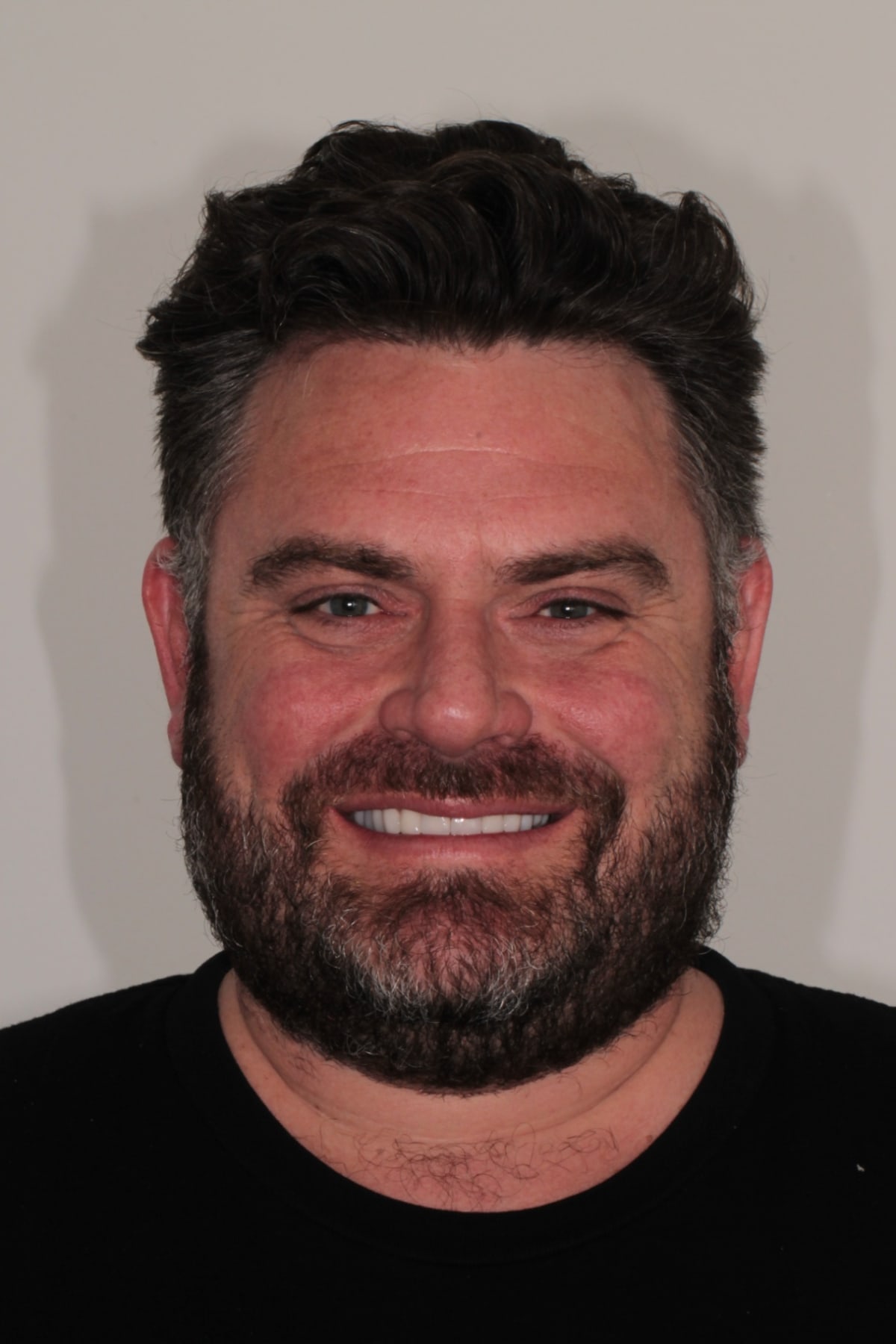
Summary:
This is a real upper full arch rehabilitation treated at ArtSmiles in Southport, now reviewed at the five year mark. The patient came to us unable to chew properly. He had lost some back teeth, the upper back teeth still in his mouth were failing, there was infection around the molars on both sides, and an upper front tooth was carrying a root canal that had failed after a childhood accident. Treatment ran for about eleven months and involved extractions, large bone grafts on both sides, six implants placed across two surgical stages, and a final set of crowns and bridges that gave him back the height of his bite. Five years on, the gums are healthy, there is no decay, and the case is stable. The honest findings are a few small chips in the back ceramic that do not affect how he chews, and one crown screw on implant 15 (upper right side) that worked loose in May 2026 and was retightened at the chair.
Key takeaways
Losing back teeth changes the front of the mouth. Without support at the back, the front teeth carry load they were never designed for and the bite gradually closes down.
Infection and missing bone come first. This case needed large bone grafts on both sides and six months of healing before a single implant could go in.
The waiting is the treatment. Two separate healing periods were built into the surgical phase on purpose.
Small technical findings at five years are normal. Published five year data on implant supported bridges reports fractures of the veneering ceramic in about 13.5% of cases and screw or abutment loosening in about 5.3%.
Six monthly reviews are what catch those findings while they are still small. This patient has not missed one.
In this article
Why this patient came to us
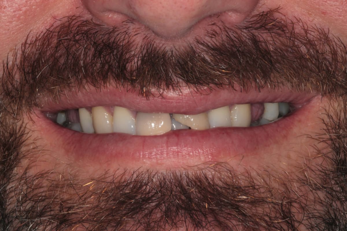
His first complaint was not about how his teeth looked. It was that he could not eat properly. He had lost several back teeth on both sides, so there was very little left to chew on, and the upper teeth that were still there were not in good condition.
The appearance mattered to him too, and he said so. There were visible gaps, and the front teeth had drifted. It is worth being blunt about what that does to someone. Missing front teeth affect how people socialise, how willing they are to speak up in a group, and how they feel about themselves. Research on tooth loss and quality of life has found the same thing repeatedly. When I sat with this patient at the first appointment, that weight was easy to see.
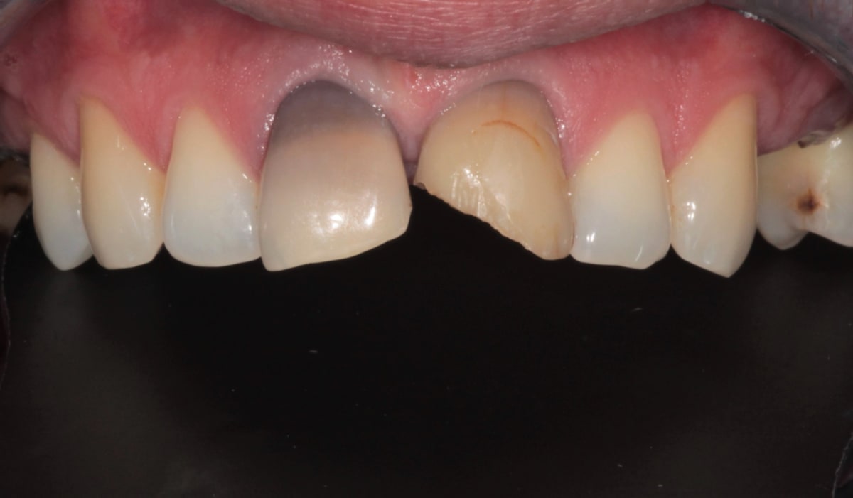
There was one more piece of history that shaped the whole plan. He had a fall as a child and a root canal was done at the time. That root canal had since failed. On the scan the canal was unusually wide, which tells you the tooth was still forming when the injury happened, so the root never finished developing properly. A tooth like that is difficult to save and even harder to keep long term.
 |  |
|---|
What the scans showed
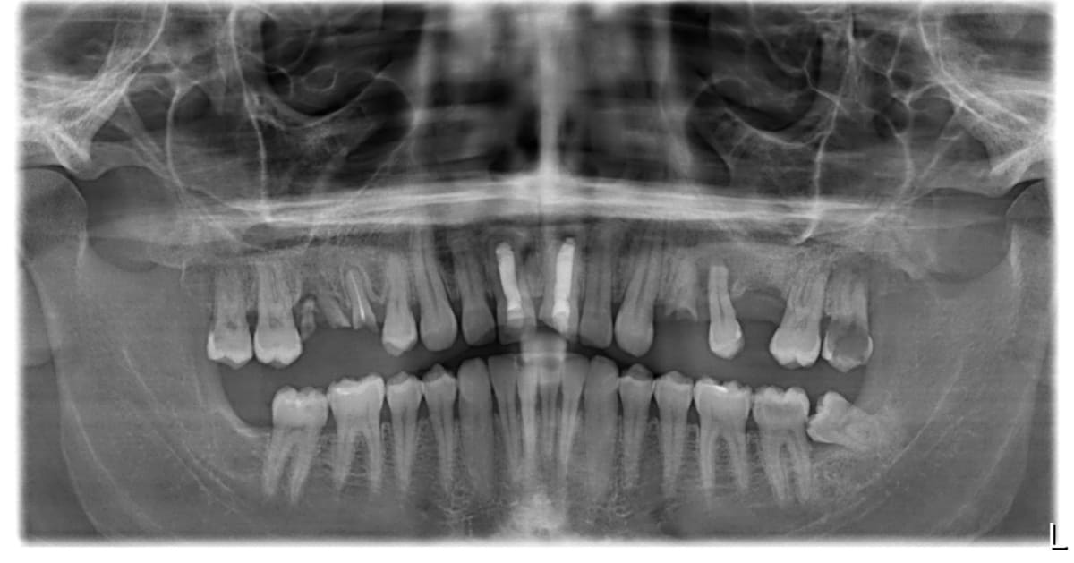
A panoramic X ray gives you the overview. A 3D scan, called a CBCT, is what lets you see the true extent of an infection and the actual volume of bone available. Both were needed here, and the 3D scan changed how I approached the case.
There was active infection around the back teeth on both sides. On one side the infection was extensive and tracked a long way up into the bone above the roots. Those teeth could not be kept, and once they came out there was going to be a large defect where healthy bone should have been. The other side had the same problem in a smaller form.
At the front, the teeth with the failed root canal showed a dark area at the tip of the root, which is what long standing infection looks like on a radiograph. Combined with the wide, underdeveloped canal, keeping that tooth would have meant a poor result that was likely to fail again. Removing it and replacing it with an implant was the more predictable option, and I said so at the planning appointment rather than trying it and hoping.
Why his front teeth had taken over the bite
This is the part of the case that is easy to miss if you only look at the gaps.
When you lose the back teeth, you lose the props that hold your bite open. The jaw closes further than it should, and the front teeth end up doing work they were never built for. In this patient the lower front teeth were biting very high, close to the gum line behind the upper front teeth. That is a deep bite, and it is a direct consequence of having nothing left at the back.
Two things follow from that. The front teeth wear and drift forward, which is exactly what had happened here. And the lower third of the face gets shorter, so the whole smile looks compressed. The literature on loss of posterior support describes this pattern well, including the flaring of the front teeth and the drop in the height of the bite.
So the plan could never be just "replace the missing teeth". It had to give him support at the back, reopen the bite to a sensible height, and only then deal with how the front teeth looked. Get that order wrong and you end up with a cosmetic result sitting on an unstable foundation.
The treatment plan
Because several problems needed attention and each one had to heal before the next step, we staged it:
Remove the infected and unsalvageable back teeth on both sides, and place large bone grafts in both areas at the same visit.
Allow six months for the grafted bone to mature.
During that waiting period, remove the two compromised upper front teeth and place two implants immediately into those sockets.
Once the grafts had matured, place the remaining implants at the back. Six implants in total.
Allow a second healing period for the implants to fuse with the bone.
Make a provisional set of teeth, assess it, correct anything it revealed, then scan and make the final crowns and bridges at the new bite height.
Finish with a custom night guard.
Total time from first appointment to the night guard was about eleven months. Almost all of that was healing, not appointments.
Stage 1: Extractions and bone grafts on both sides
The first surgical visit dealt with the infection. The compromised back teeth came out on both sides, the sockets and the surrounding infected tissue were thoroughly cleaned, and bone graft material was placed into both defects to rebuild the ridge.
A bone graft is scaffolding. It gives your own bone a framework to grow into, and over several months the graft is gradually replaced by living bone that an implant can anchor into. It is not instant, and it cannot be rushed. A systematic review by Aghaloo and colleagues (2016) found that bone augmentation in the upper jaw reliably creates enough bone for implant placement, which is why this step is worth the wait rather than something to skip.
I waited a full six months before touching those areas again. That was a deliberate choice. Placing an implant into a graft that has not fully matured is how you get an implant that never integrates properly.
Stage 2: Two immediate implants at the front
Six months of waiting is a long time to leave someone with failing front teeth, so we used that window rather than sitting still.
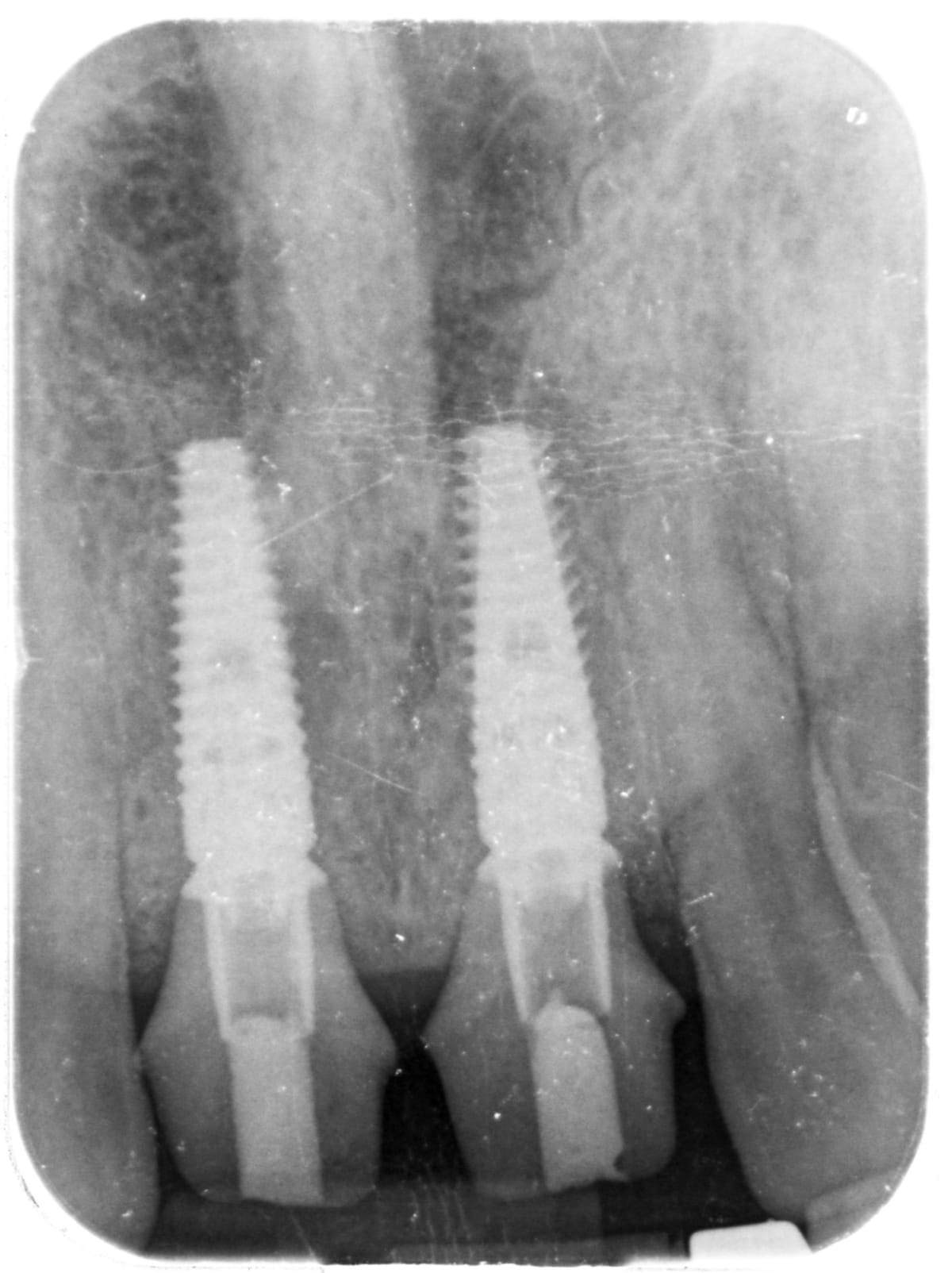
While the back grafts were maturing, I removed the two upper front teeth, including the one with the failed childhood root canal, and placed two implants into those sockets at the same appointment. That is called immediate implant placement. Both sites had small areas of infection at the root tips, which used to be considered a reason not to place an implant straight away.
The evidence has moved on. A 2024 systematic review and meta analysis comparing immediate implants placed into sockets with and without infection at the root tip found survival rates that were essentially the same between the two groups, provided the socket is properly cleaned and debrided first. That last part is the whole game. The implant does not fail because there was infection. It fails if the infection is still there when you place it.
Stage 3: The back implants and the second healing period
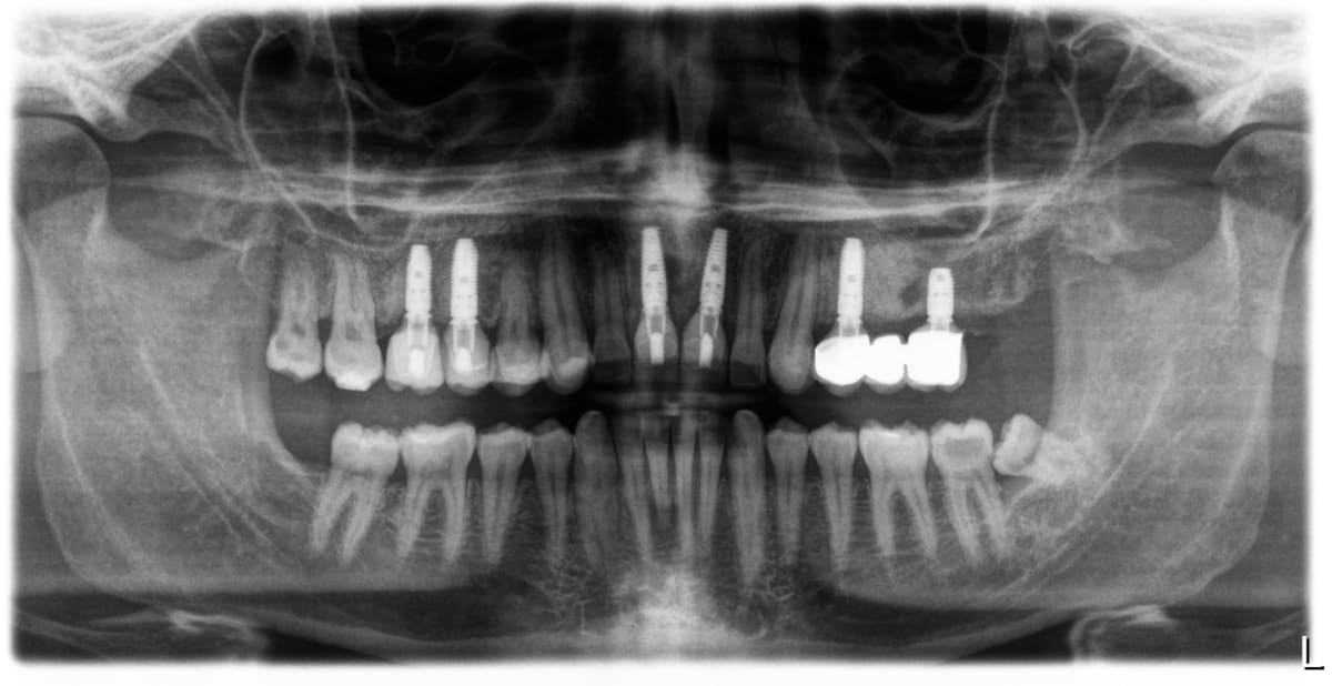
Once the grafts had matured into solid bone, I went back and placed the remaining implants at the back, bringing the total to six across the upper arch.
Then came the second waiting period. Implants work because bone grows directly onto the titanium surface and locks it in place, a process called osseointegration. Nothing you do speeds that up. You place the implant correctly, you keep the area clean, and you let biology do its part.
Long term data is what makes this wait easy to justify to a patient. A systematic review by Moraschini and colleagues (2015), pooling more than 7,700 implants with an average follow up of over 13 years, reported implant survival of 96.4% at ten years with minimal bone loss around them. That is the payoff for staging the surgery properly.
Stage 4: The provisional, and the problem it found
Before any final crown gets made, we make a provisional set. It is quick, it is made in the chair, and it is not meant to be perfect. Its job is to test the plan in the patient's mouth, at the new bite height, so that any problem shows up while it is still cheap and easy to fix.
It earned its keep here. When I photographed the provisional and looked at it properly, one side of the arch was not filling out the corner of the smile the way the other side was. The technical term is the buccal corridor, which is the dark space you see between the back teeth and the corner of the mouth when someone smiles widely. Too much of that space on one side and the smile looks lopsided even when every tooth is technically in the right place.
This happens for ordinary reasons. Sometimes the mould shifts slightly when a chairside provisional is being made. Sometimes it is simply hard for a laboratory to be perfectly accurate across that many teeth at once. Either way, the fix was straightforward. I added composite in the chair so he could bite and function comfortably, and I sent the photographs to the laboratory with a clear instruction to build that area out on the definitive restorations.
That is the honest reason we use provisionals. Not because we expect them to be perfect, but because they show you what to change before you commit to porcelain.
Stage 5: The final crowns and bridges
With the implants integrated and the provisional corrected, I scanned the mouth digitally and the laboratory made the definitive restorations.
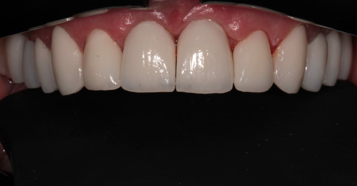
The final work combined a bridge supported by two implants, crowns on the two front implants, veneers on other upper teeth and single crowns on the remaining implants. Everything was checked for stability before anything was fitted permanently. The restorations were built to the corrected bite height, so his back teeth carried the load again and his front teeth went back to doing what front teeth are supposed to do, which is guide the jaw and look natural.
If you want more detail on how implant supported crowns and bridges are put together, or on single implants for individual missing teeth, those pages walk through the process.
 |  |
|---|
Try Smile Studio
Curious how a new smile might look on you before you commit to anything? Try Smile Studio, our free online tool. Upload a photo of your smile and preview different tooth shapes and shades right in your browser, in your own time and with no pressure. It works best on a computer or laptop, so open it on a desktop rather than your phone for the easiest experience. It is a simple way to explore the look you are after and bring those ideas to your consultation, so we can talk through what is realistic for your teeth. Keep in mind it is a visual guide to spark the conversation, not a treatment plan or a promise of results.
The night guard at the end
The last appointment of the treatment was the night guard, and it is not an afterthought.
This patient grinds. On a case with this much ceramic in it, that matters, because grinding forces concentrate on the hardest surfaces in the mouth, and after this treatment the hardest surfaces were his new restorations. A custom guard spreads that load across a piece of acrylic instead of across the porcelain.
We finish every large rehabilitation this way, whether or not the patient has a known grinding habit. It is inexpensive insurance on a case that took eleven months to build. You can read more about how we manage grinding on our bruxism treatment page.
The five year review
He has come back every six months since the case was finished, and has not missed a review. That consistency is a big part of why this section reads the way it does.
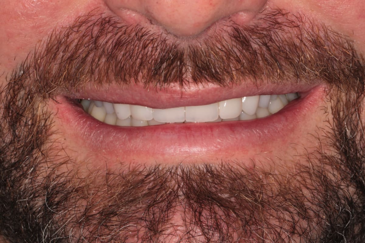
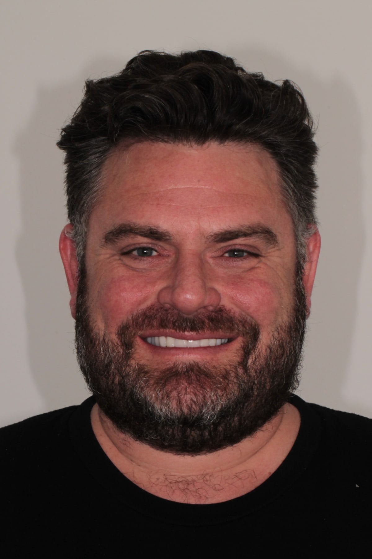
The good news first. The gum tissue around the implants is in excellent condition, with no inflammation and no bleeding. There is no decay anywhere in the mouth. The bite is stable, the implants are stable, and the restorations are doing the job they were made for. He is pleased with how his teeth look and, more to the point for him, he can eat normally.
Now the two honest findings.
First, there are a few small chips in the ceramic on the back teeth. They are minor, they do not affect how he chews, and they have not required replacing anything. This is the most common technical finding in implant supported bridges. In Pjetursson and colleagues' systematic review (2012), fractures of the veneering ceramic occurred in about 13.5% of cases over five years, which was the single most frequent complication reported. Seeing small chips at year five is not a sign that something went wrong. It is the expected behaviour of ceramic under years of grinding load.
Second, in May 2026 the crown on implant 15, the upper right second premolar, came loose. What loosens in a case like this is almost never the implant. It is the small screw that holds the crown onto the implant, which can gradually back off under repeated chewing and grinding forces. He came in, I removed the crown, checked the implant and the fitting surfaces, and retightened the screw to the correct torque. There was no damage to the crown and no damage to the implant, and he left the same appointment with it back in function. The same review put screw or abutment loosening at about 5.3% over five years.
Both of those findings are the reason I keep telling patients that a rehabilitation is not a finished object. It is something you maintain. A systematic review by Monje and colleagues (2016) found roughly a 25% lower incidence of peri-implantitis in patients on a regular maintenance programme, and suggested a review interval of five to six months. This patient's six monthly schedule is exactly why a loose screw was a fifteen minute appointment rather than a fractured crown.
Before | After | After 5 years |
|---|---|---|
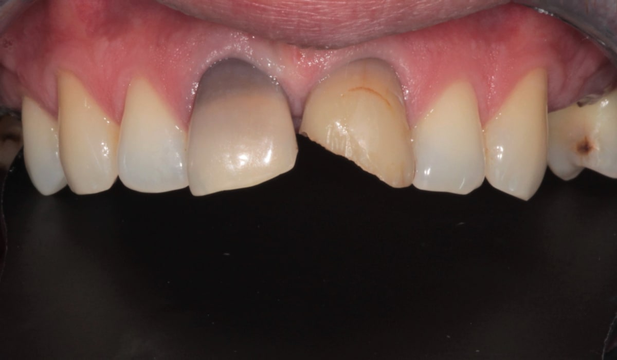 | 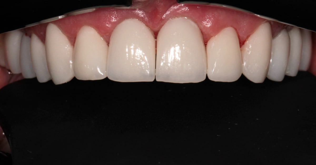 | 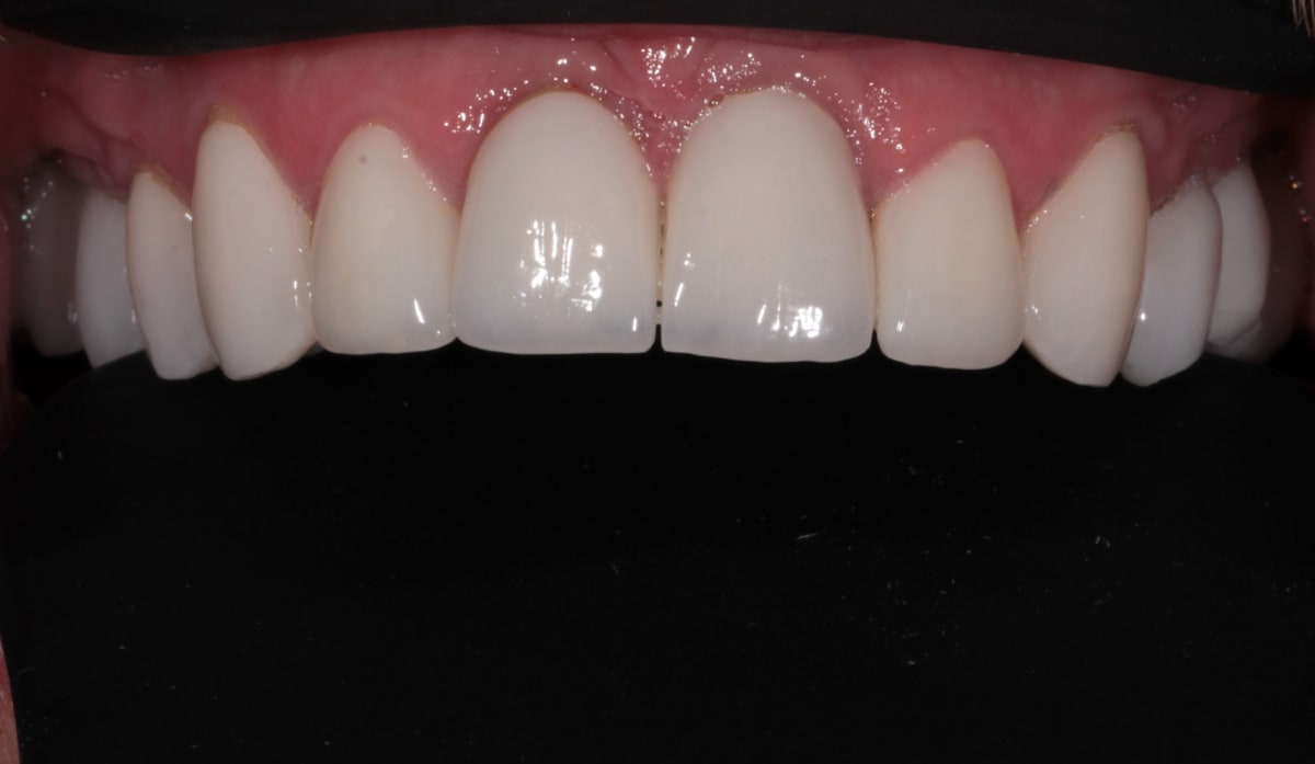 |
After | After 5 years |
|---|---|
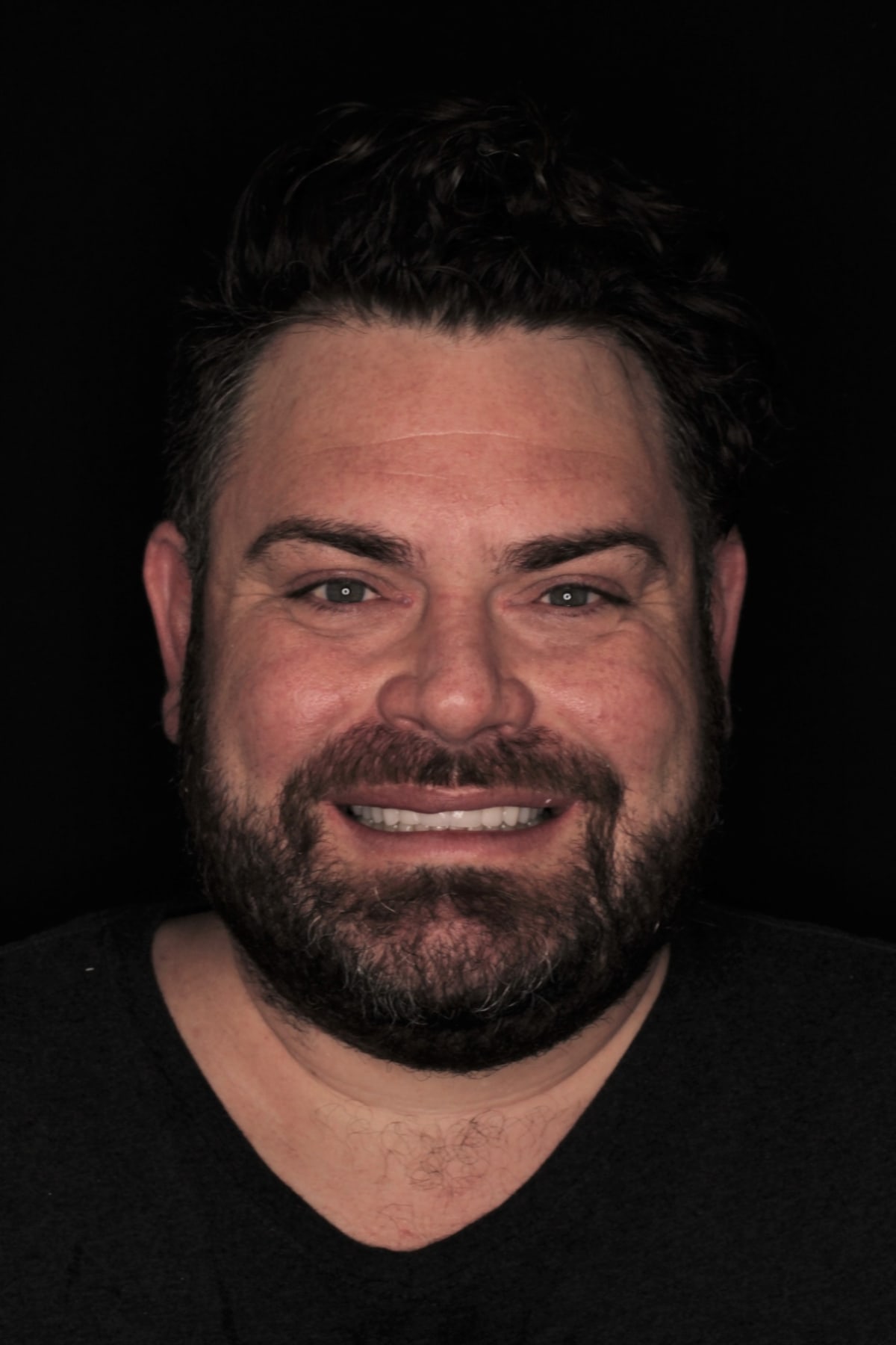 |  |
What this case tells us at five years
Looking back through the file, four things stand out.
The sequence mattered more than any single procedure. Infection first, then bone, then implants, then the bite, then the appearance. Every part of this case that has held well for five years can be traced back to something that was allowed to heal properly before the next step started.
Reopening the bite was not cosmetic. It was structural. Giving him back support at the back is what stopped his front teeth carrying a load they were not designed for, and that is why the front of the case still looks the way it did on the day it was fitted.
The provisional was worth every minute. The buccal corridor problem would have been an expensive thing to discover in porcelain. In acrylic it was a conversation with the laboratory and a bit of composite.
And the small problems are not failures. A few ceramic chips and one loose screw over five years, both caught at a routine review and both fixed without replacing anything, is a case behaving normally. What would worry me is a patient who disappears for five years and turns up with the same two findings after they have had time to become something bigger. You can see how other long term cases have held up in our clinical case gallery, including a rebuild after severe gum disease, and our guide to how long dental implants last covers what drives longevity in more detail.
Disclaimer
This article documents one patient's treatment at ArtSmiles. It is shared for educational purposes with the patient's written consent. Individual results vary and depend on factors including oral health, bone and gum condition, general medical history, and how well the restoration is maintained after treatment. Nothing in this article is a guarantee of outcome, a substitute for a clinical examination, or advice specific to your case. Any treatment carries risks and potential complications, which will be explained to you at consultation.
Before starting any dental treatment, book a consultation so we can assess your teeth, gums, and bone in person, take the imaging we need, and discuss the options, timelines, and costs that apply to your situation.
Case executed by Dr Cristian Dunker
General Dentist
AHPRA DEN0002257085
ArtSmiles, Southport, Gold Coast
Frequently asked questions
How long does a full upper rehabilitation with implants take?
This case took about eleven months from the first appointment to the final night guard. Most of that time was healing rather than treatment. The bone grafts needed six months to mature before implants could be placed, and the implants then needed their own healing period to fuse with the bone. If bone grafting is not required, the timeline is usually shorter. If more grafting is needed, it can be longer. Your dentist should be able to give you a realistic range after a 3D scan, not before one.
Why do I need a bone graft before dental implants?
An implant needs a solid volume of bone to anchor into. When teeth are lost to infection or gum disease, the bone that held them often goes with them, so there may not be enough left to place an implant safely. A bone graft rebuilds that volume. It acts as a scaffold that your own bone grows into over several months, and once it has matured it can support an implant. Skipping this step to save time is one of the more common reasons implants fail later.
Can an implant be placed straight after a tooth is removed if there is infection?
Often yes, provided the socket is thoroughly cleaned and debrided at the same visit. That was the approach used at the front of this case. Published evidence comparing immediate implants placed into sockets with and without infection at the root tip has found comparable survival rates when the site is properly cleaned first. It still needs case by case judgement, and there are situations where waiting is the better call, which is why it is a decision made with a 3D scan in front of you.
What happens if a crown on an implant becomes loose?
In most cases it is the small screw holding the crown to the implant that has worked loose, not the implant itself. It is a straightforward fix. The crown is removed, the implant and fitting surfaces are checked, and the screw is retightened to the correct torque. That is exactly what happened with implant 15 in this case in May 2026, with no damage to either the crown or the implant. The important part is coming in promptly. Leaving a loose crown in function is what turns a simple retighten into a fractured restoration.
Are chips in the ceramic normal after five years?
Small chips in the ceramic on the back teeth are the most commonly reported technical finding in implant supported bridges, occurring in roughly 13.5% of cases over five years in published reviews. Minor chips that do not affect function are usually polished and monitored rather than replaced. They are more likely in patients who grind, which is why a night guard is part of the treatment and not an optional extra.
How often should I be reviewed after implant treatment?
Every six months for most patients. Research on peri-implant maintenance suggests an interval of five to six months, and found around a 25% lower rate of peri-implantitis in patients who attend regularly compared with those who do not. Reviews are where small things get found while they are still small. A loose screw picked up at a routine visit is a short appointment. The same screw left for a year is a different conversation.
If you are struggling to chew, living with missing back teeth, or have been told your upper teeth cannot be saved, we offer a complimentary implant consultation where we can look at your scans and talk through what a staged plan would realistically involve for you. There is no obligation to proceed. You can also read more about full mouth rehabilitation and missing teeth.
References
Aghaloo et al. (2016). Bone augmentation of the edentulous maxilla for implant placement: a systematic review. Int J Oral Maxillofac Implants. PubMed
Moraschini et al. (2015). Evaluation of survival and success rates of dental implants reported in longitudinal studies with a follow-up period of at least 10 years: a systematic review. Int J Oral Maxillofac Surg. PubMed
Pjetursson et al. (2012). A systematic review of the survival and complication rates of implant-supported fixed dental prostheses (FDPs) after a mean observation period of at least 5 years. Clin Oral Implants Res. PubMed
Monje et al. (2016). Impact of maintenance therapy for the prevention of peri-implant diseases: a systematic review and meta-analysis. J Dent Res. PubMed
Pranckeviciene et al. (2024). Comparison of immediate implantation into the socket with and without periapical pathology: a systematic review and meta-analysis. Medicina (Kaunas). PubMed Central
Craddock et al. (2007). Occlusal changes following posterior tooth loss in adults. Part 2. Clinical parameters associated with movement of teeth adjacent to the site of posterior tooth loss. J Prosthodont. PubMed
Written by Dr Cristian Dunker, principal dentist at ArtSmiles Cosmetic Dentistry, Southport.
Medically reviewed on 4 August 2026 by Dr Cristian Dunker.