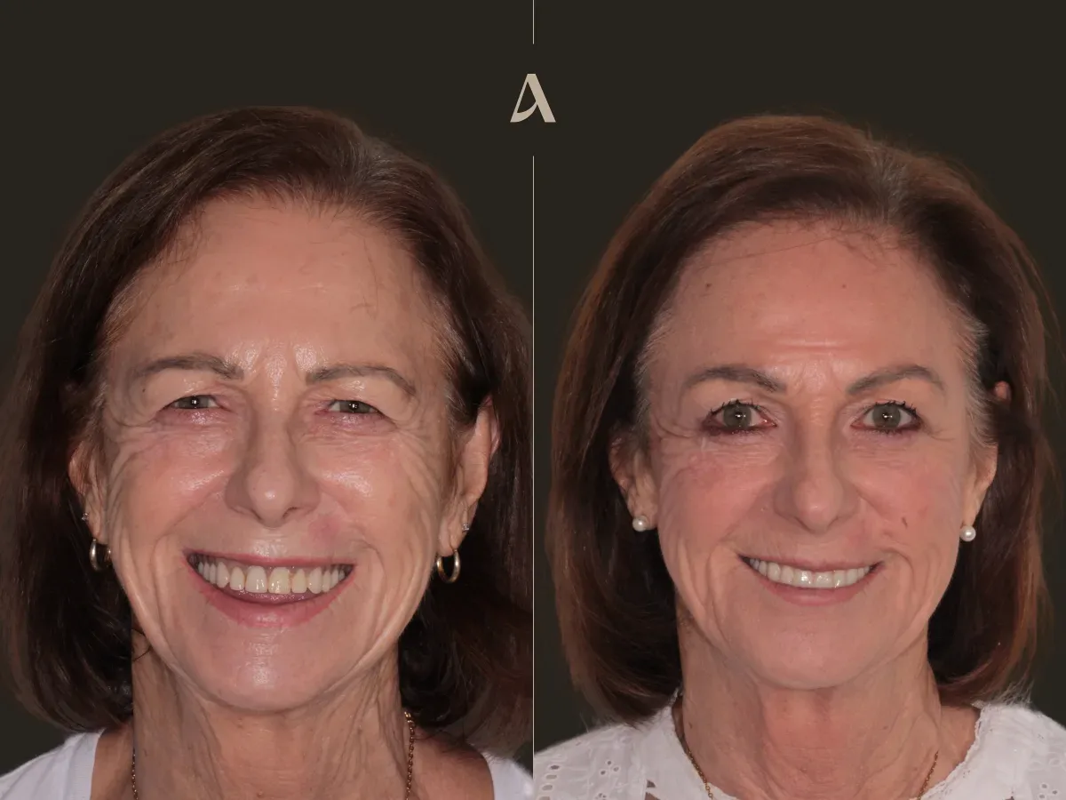
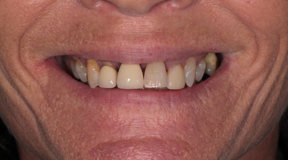
This patient came to ArtSmiles wanting to fix her upper teeth. She was unhappy with how her front teeth looked and had been living with discomfort for some time. What started as a worry about appearance turned out to be a deeper problem, and putting it right meant rebuilding her upper smile from the foundation up.
This is a real clinical case treated at our Southport practice on the Gold Coast. It shows how severe gum disease in the upper front teeth, combined with a bite that had gradually closed down over the years, can be rebuilt into a stable and natural looking smile using dental implants and zirconia. From the first appointment to the final result, the treatment took twelve months.
Losing front teeth is rarely just about appearance. Research consistently links anterior tooth loss to a real impact on confidence and quality of life (Gerritsen et al., 2010), and I could see that weight on this patient from our first conversation. Walking through how we planned and staged her treatment may help anyone facing something similar understand what is possible and why each step matters.
In this article
What I Found During the Examination
A careful clinical examination and a 3D X ray, also called a CBCT scan, gave me a complete view of what was happening beneath the surface. There were two main problems.
First, there was severe gum disease around the upper front teeth. Advanced periodontal disease had destroyed much of the bone that held the four upper front teeth in place. These teeth were failing, mobile, too inclined and could not be saved.
One of these teeth, the upper right central incisor (tooth 11), had an old post inside the root, a small rod used to support a previous restoration. On the scan it also showed a periapical radiolucency, a dark area at the very tip of the root that points to a long standing infection. A post like this makes a tooth more fragile, and with infection sitting at the root tip as well, this tooth was especially difficult to save.
Second, her bite had collapsed over the years, a problem we call reduced vertical dimension. When the bite loses height, the smile looks shorter, the lower face can appear to fold in a little, and the remaining teeth take on more strain than they were built for.
The same disease that had loosened the front teeth had also worn away the bone in that area, so the ridge would need to be rebuilt before any implants could be placed. The back teeth and the lower arch were in a condition we could work with, so my focus was on rebuilding the upper front and restoring the bite.
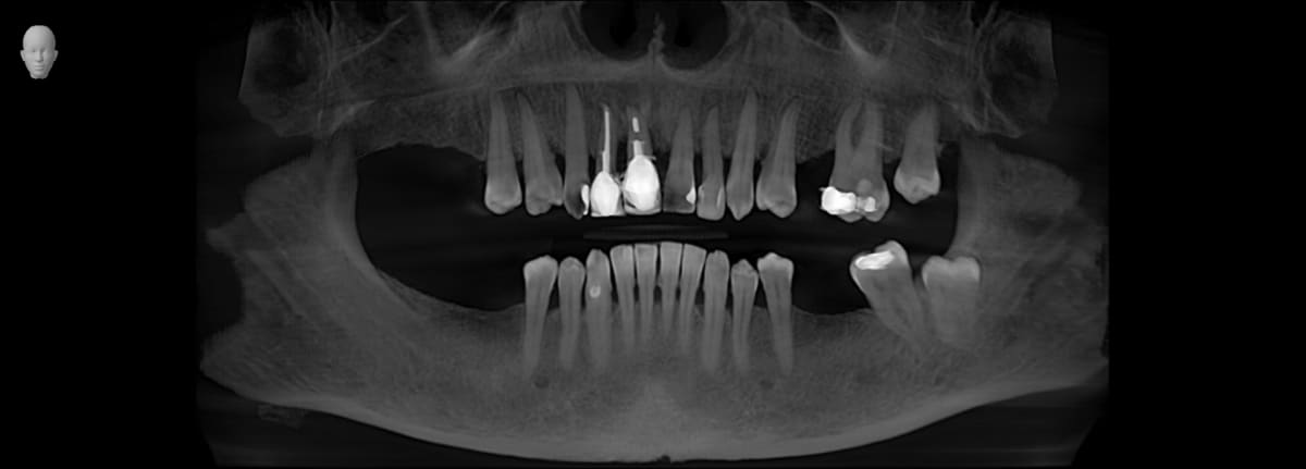
This image shows large amount of tartar in both arches, moderate to severe bone loss caused by gum disease and severe bone destruction around upper incisors (forth and fifth teeth upper from left to right) and several missing teeth.
 |  |
|---|
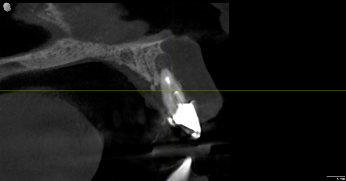
These two x-ray slices show different problems in the same area. The darkness around tooth 11 (central incisor) shows long term infection created by two different problems: gum disease and failed root canal. Unfortunately the best restorative result would require extraction of the affected teeth.
 |  |
|---|
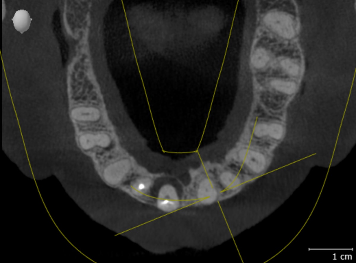
Preoperative images show front teeth more angulated than normal, caused by absence of back support and severe gum disease in front teeth. The treatment must consider realigning the front teeth to provide better aesthetics.
The Treatment Plan
Because several things needed attention at once, we planned a staged full mouth rehabilitation. Treating the gum disease first, then rebuilding the bone, then placing the implants, and finally restoring the smile and the bite gives each step time to heal properly before the next one begins. The plan looked like this:
Treat the active gum disease and remove the four upper front teeth that could not be saved.
Place a bone graft in the upper front area to rebuild the lost ridge.
Fit a fixed temporary bridge across the front so she was never without teeth while everything healed.
Place dental implants once the bone had matured, two to support the new upper front teeth and one to replace a missing tooth in the lower left.
Rebuild the smile in zirconia (upper and lower arches) and reopen the bite to its natural height.
Treating the gum disease before anything else was not optional. As Sousa et al. (2016) showed in their systematic review, patients with a history of periodontitis face higher rates of complications around implants, so settling the gums first is one of the most important steps in giving the implants the best chance of lasting.
Stage 1: Treating the Gum Disease and Rebuilding the Foundation
We began with a thorough deep clean to bring the active infection under control. Once the gums had settled and the inflammation had reduced, I removed the four upper front teeth that could not be saved.
In the same visit, I placed a bone graft into the area where the teeth had been. A healthy volume of bone is what gives an implant something solid to anchor into, so rebuilding the ridge now is what makes a strong result possible later. To make sure she left with a complete smile, we fitted a fixed temporary bridge spanning from one canine to the other, which she wore while the graft healed underneath.
 |  |
|---|
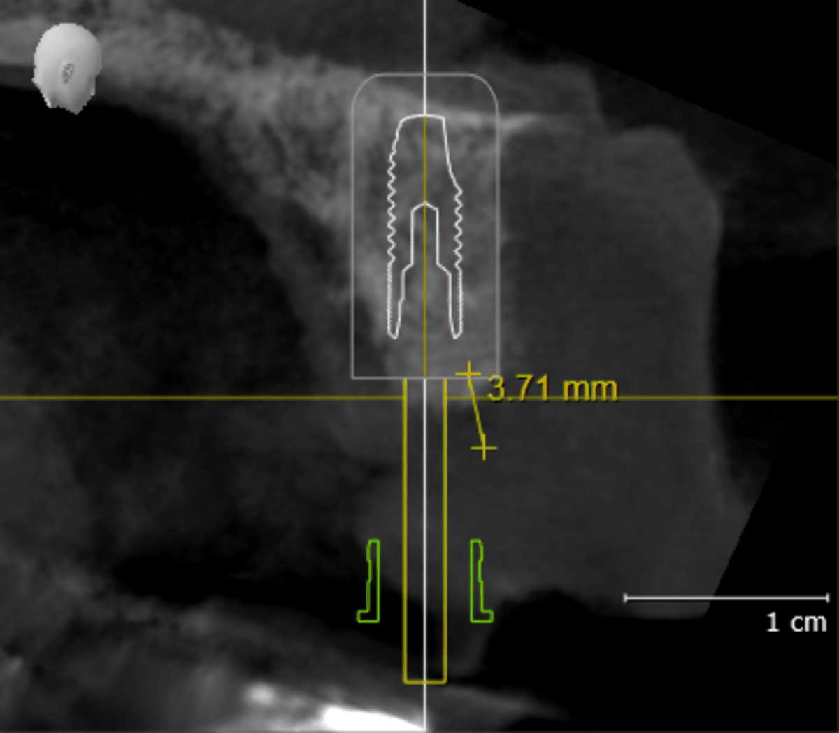
These two x-ray slices show the same area. On the left, the tooth with severe bone loss and infection, and on the right, the same area shows bone graft after healing, during the planning for implants. You can notice large gain of bone volume with bone graft, and implant planning with a better implant angulation.
Try Smile Studio
Curious how a new smile might look on you before you commit to anything? Try Smile Studio, our free online tool. Upload a photo of your smile and preview different tooth shapes and shades right in your browser, in your own time and with no pressure. It works best on a computer or laptop, so open it on a desktop rather than your phone for the easiest experience. It is a simple way to explore the look you are after and bring those ideas to your consultation, so we can talk through what is realistic for your teeth. Keep in mind it is a visual guide to spark the conversation, not a treatment plan or a promise of results.
Stage 2: Placing the Implants
After several months, once the graft had matured into solid bone, I placed three implants. Two went into the upper front to support the new front teeth, and one went into the lower left to replace a missing back tooth. The implants were then left to integrate with the bone, a natural healing process called osseointegration that gives implants their strength.
A systematic review by Aghaloo et al. (2016) confirmed that bone grafting in the upper jaw can successfully rebuild the ridge for implant placement with high success rates. Research by Hof et al. (2015) looking at the timing of implants in the front of the mouth found that placing them after the graft has healed achieves reliable long term results. Throughout this healing time she kept wearing her temporary bridge, so her appearance and confidence were never interrupted.
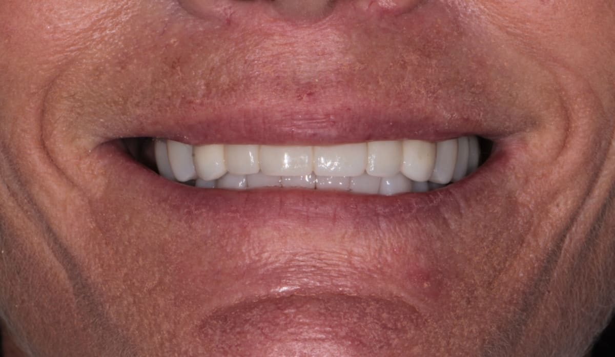
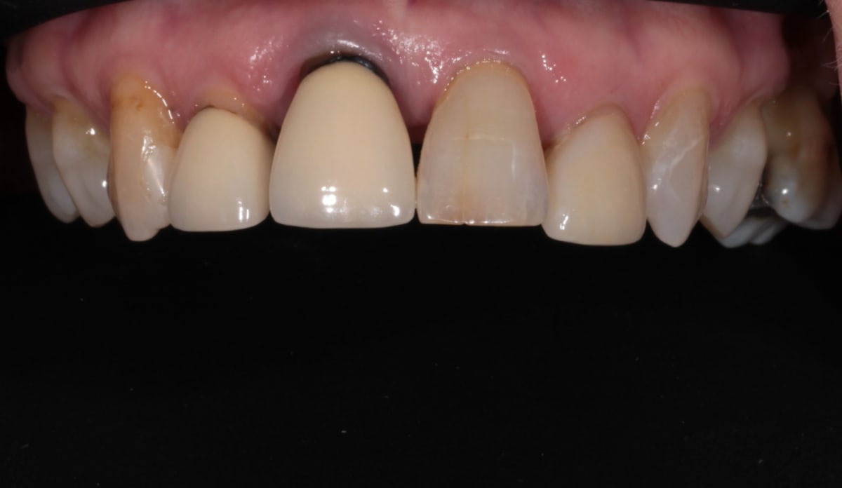
Smile with long term temporary bridge. The patient wore this bridge for 9 months.
Stage 3: The Final Zirconia Smile
With the implants fully healed, we moved on to the part she had been waiting for. Working with our dental laboratory, 3Dent, we rebuilt her smile in zirconia, a strong material that can be layered to look natural. This included zirconia veneers and crowns on her own teeth (upper and lower arches), a zirconia bridge fixed onto the two front implants, and a crown on the lower implant.
Just as importantly, we used these restorations to reopen her bite to its proper height and restore the vertical dimension that had been lost over the years. This made her teeth look more youthful, and it gave her jaw muscles a more comfortable and balanced position to work from.
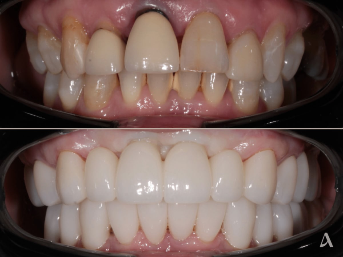
The Result
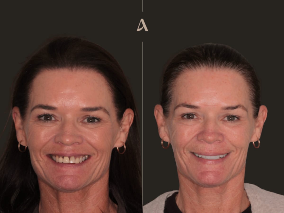
Twelve months after her first visit, she had a fixed, stable upper smile that looked and felt like her own teeth. The infection was gone, the bone was rebuilt, the bite was restored to its natural height, and her front teeth were now supported by implants rather than failing roots.
Research by Ramani et al. (2020) found that implant supported fixed restorations in the smile zone have a strongly positive effect on quality of life, with patients reporting high satisfaction with how their teeth look, feel, and function. A study by Chen et al. (2012) similarly showed that replacing missing front teeth with implants improves dental confidence. For a patient who had walked in feeling self conscious, the change in how she carried herself was the most rewarding part of the whole case.
 |
|---|
Key Takeaways from This Case
Gum disease in the front teeth is not just a cosmetic issue. It affects the bone, the bite, and the long term health of the whole mouth.
Bone often needs rebuilding first. When teeth are lost to gum disease, a bone graft is usually needed before implants can be placed.
A staged approach is worth the wait. Letting each part heal properly is what gives a result like this its strength.
You are never left without teeth. A fixed temporary bridge keeps you smiling and eating throughout the healing.
Restoring the bite matters as much as replacing teeth. Reopening a collapsed bite is often the key to a result that looks natural and feels comfortable.
If you are living with loose front teeth, gum disease, or a bite that has worn down over the years, I would be glad to help. Book a consultation at ArtSmiles and we can talk through what is possible for your smile.
Disclaimer
This article documents one patient's treatment at ArtSmiles. It is shared for educational purposes with the patient's written consent. Individual results vary and depend on factors including oral health, bone and gum condition, general medical history, and how well the restoration is maintained after treatment. Nothing in this article is a guarantee of outcome, a substitute for a clinical examination, or advice specific to your case. Any treatment carries risks and potential complications, which will be explained to you at consultation.
Case executed by Dr Cristian Dunker
General Dentist
AHPRA DEN0002257085
ArtSmiles, Southport, Gold Coast
Medically reviewed by Dr Cristian Dunker.
Frequently Asked Questions
Can you have dental implants if you have gum disease?
Yes, but the gum disease has to be treated and brought under control first. Implants need healthy gums and solid bone to succeed. In this case we settled the periodontitis with a deep clean before planning any implants, which research by Sousa et al. (2016) supports as an important step in lowering the risk of complications.
Will I be left without front teeth during treatment?
No. This patient wore a fixed temporary bridge across her front teeth for the whole healing period, so she always had a complete smile while the bone and implants healed underneath.
Why did the bite need to be reopened?
Years of wear and tooth loss had closed her bite down, which we call reduced vertical dimension. Rebuilding the teeth to their proper height makes the smile look more youthful and gives the jaw muscles a more comfortable position, so it is an important part of a full mouth rehabilitation.
Why use zirconia for the veneers, crowns, and bridge?
Zirconia is very strong and resists chipping, while still looking natural because it can be layered to mimic real teeth. That mix of strength and appearance makes it a good choice across veneers, crowns, and a bridge fixed onto implants.
How long does this kind of treatment take?
This case took twelve months from the first appointment to the final restoration. The timeline is driven by healing, because the bone needs time to mature after grafting and the implants need time to fuse with the bone before the final teeth go on.
References
Gerritsen et al. (2010). Tooth loss and oral health-related quality of life: a systematic review and meta-analysis. Health Qual Life Outcomes. PubMed
Sousa et al. (2016). A systematic review of implant outcomes in treated periodontitis patients. Clin Oral Implants Res. PubMed
Aghaloo et al. (2016). Bone augmentation of the edentulous maxilla for implant placement: a systematic review. Int J Oral Maxillofac Implants. PubMed
Hof et al. (2015). Does timing of implant placement affect implant therapy outcome in the aesthetic zone? Clin Implant Dent Relat Res. PubMed
Ramani et al. (2020). Patient satisfaction with esthetics, phonetics, and function following implant-supported fixed restorative treatment in the esthetic zone. J Esthet Restor Dent. PubMed
Chen et al. (2012). The psychosocial impacts of implantation on the dental aesthetics of missing anterior teeth patients. Br Dent J. PubMed